ASSESSMENT OF ULTRAVIOLET RADIATION EXPOSURES IN
PHOTOBIOLOGICAL EXPERIMENTS
J.C.F. Wong1 and A.V. Parisi2
1Centre for Medical and Health Physics, School of Physical Sciences, Queensland University of Technology, GPO Box 2434, Brisbane, 4001, Australia.
Ph. +61 7 3864 2585. FAX: +61 7 38641521
2
Centre for Astronomy and Atmospheric Research, Department of Biological and Physical Sciences, University of Southern Queensland, Toowoomba, 4350, Australia. Ph: +61 7 46 312226. Fax: +61 7 46 312721.ABSTRACT
The interfering effect of ultraviolet (UV) radiation on the natural function of biological processes is wavelength specific and the UV spectrum must be weighted with the action spectrum for the process. The UV spectral irradiance may be measured with calibrated spectroradiometers. Alternatively, the biologically effective UV may be measured with broadband devices. This paper reviews the techniques for assessing biologically effective exposures in photobiological experiments.
UV meters, such as the Robertson-Berger (RB) meter, or passive dosimeters, such as polysulphone, that possess a spectral response approximating the human erythemal response can be used to estimate erythemally effective exposure or actinic exposure due to solar UV. The sensitivity of the RB meter is about 0.56 m W cm-2 and polysulphone can record an exposure of about 2 mJ cm-2. For photobiological processes other than erythema these devices are not suitable to determine the exposure. In terms of these applications, a spectrum evaluator consisting of four different types of dosimeter material can be employed to evaluate the UV spectrum of the source. This method can be useful both for solar UV studies and research with UV lamps that possess radiation wavelengths shorter than 295 nm. The device can be used to measure exposures where the actinic and erythemal action spectra differ significantly. It can also be used to assess exposure due to low levels of UV (about 0.01 m W cm-2) caused by radiation filtered through glasses or plastic.
TABLE OF CONTENTS
1. Background
2. Photobiological Processes
3. Biologically Effective Irradiance
4. Spectroradiometry
5. Broadband Measurements
6. Spectrum Evaluator
Practicality
Conclusion
1. Background
Ultraviolet radiation (UVR) exists in the environment due to the natural source of the sun and/or artificial light sources. On the surface of the earth, the wavelengths of the solar UV radiation lies mainly in the UVB waveband (290 nm – 320 nm) and the UVA wavebands (320 nm – 400 nm). The waveband of artificial light sources may extend from the UVC waveband (200 nm – 400 nm) to the UVA waveband depending on the type of the source. The effect of UV radiation on a biological specimen is determined by the spectral irradiance, El , delivered to the surface of the biological body and the duration of the exposure, T. The total exposure, E, is a time integrated function of the spectral irradiance
![]()
Because the spectral irradiance may vary with the time and the site of exposure, it is a complicated function of these two variables. To determine the spectral irradiance, a calibrated spectroradiometer (Wong et al., 1995) can be used. A sensor sensitive to a selected UV waveband may be calibrated against a spectroradiometer (Wong et al., 1995) for broadband measurements. For various applications, the broadband meters may be classified as the UVA meter, the UVB meter or the UVR meter. The time integration may be carried out electronically so the instrument can provide a display of the total irradiance. The total irradiance can also be determined using a passive dosimeter. A material sensitive to a selected waveband is useful for the purpose. It must be calibrated against other instruments prior to usage.
The total irradiance (J m-2) provides information about the total photon energy (joules, J) in the UV waveband falling on a unit area (meter2, m2) of the surface of the body in consideration. Because the response of the biological body to UVR varies with the wavelength of the radiation, this quantity is not a good index of biological effects. To assess the biologically effective exposure to a selected biological tissue, it is necessary to know the sensitivity of the tissue to UVR. This method requires rescaling the spectral irradiance by the relative sensitivity of the biological tissue. It will be described in detail in section 3. A broadband UV meter or a passive dosimeter can be used for the measurement of the biologically effective exposure. The sensor should have a spectral response suitable for the assessment of the effect of UVR on the biological body in consideration.
2. Photobiological Processes
2.1 Introduction
The biological effects of UVR may be classified into three categories: the effect on a cell, the effect on a tissue, and the effect on the whole body. At the cellular level, the most important effect is the DNA alteration due to the absorption of photons in the UVR waveband. For examples, the formation of single-strand breaks (SSBs), DNA to protein crosslinks (DPCs) and double-strand breaks (DSBs) have been observed in both animal and human cells in culture (Peak and Peak, 1991, Churchill et al., 1991). They concluded that UVR at around 365 nm is at least one order of magnitude more efficient to cause the lesion than ionising radiation. Exposure of cells to UVR can also affect the base substances, such as pyrimidine, in the DNA polymer in situ. The formation of pyrimidine dimer in human skin, in situ, by UV exposure has been reported by a number of authors (Freeman et al., 1987, Hacham et al., 1990). Repair mechanisms can take place in the cell for UV damage (Holmberg et al., 1985, Roza et al., 1985, Sharma and Smith, 1985). Incomplete recovery may lead to the mutation of the cell or the killing of the cell.
In a biological tissue, these processes are complicated by the presence of factors such as oxygen (Tyrrell and Pidoux, 1989). Cellular damage by UVR may cause a change in the appearance of tissue such as the induction of erythema and the ageing of the human skin (Longstreth et al., 1995).
In terms of the effect of UVR to the whole body, the damage can cause both physiological effects and impairment of the body function. In the case of animals and humans, the effect of UVR can lead to skin cancers, eye disorder and immuno-suppression (Longstreth et al., 1995). On the other hand, the exposure to UVR can affect the plant growth and photosynthesis (Caldwell et al., 1995).
2.2 Action Spectra
For a given photobiological process, the wavelength dependence of the relative spectral effectiveness, Sl , is the action spectrum. Coohill (1989) and (1991) review the importance of action spectra in photobiology. Caldwell et al. (1983) provided the action spectrum for DNA damage (Figure 1) by Setlow (1974) as:
Sl = e13.82(1.0/D-1.0) where D = 1.0+e[(l -310)/9] (2.1)
where the wavelength is in units of nm. The relative effectiveness for producing UV damage on humans decreases by about four decades over a waveband between 280 nm and 320 nm. This action spectrum has been employed for DNA damage in both humans and plants. In addition to this action spectrum for DNA damage, there exists an action spectrum for DNA damage in intact alfalfa seedlings (Quaite et al., 1992). The two are similar for wavelengths longer than 310 nm with the latter being less sensitive to UV at shorter wavelengths.
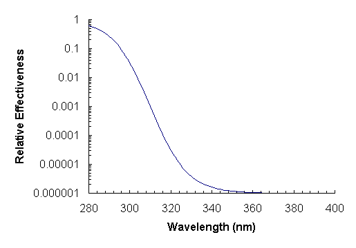
Figure 1 - Action spectra for DNA damage (Setlow, 1974).
The CIE (1987) erythemal action spectrum for humans (Figure 2) has been employed widely for assessing the UV effect on human skin. The value of the sensitivity is normalised to unity at 298 nm and drops by about 3 decades up to 320 nm. For longer wavelengths, 320-400 nm, the value of the action spectrum varies from 10-3 to 10-4. Anders et al. (1995) re-evaluated the erythemal action spectrum with a tunable dye laser and found a second maximum of lower relative response than the first maximum at 298 nm in the UVA waveband at 362 nm. The action spectrum for skin cancer induction in hairless mice (de Gruijl et al., 1993) also contains a second maximum in the UVA at approximately 380 nm. The IRPA (1989) actinic action spectrum (Figure 2) has been employed for assessing the UV effects on both human skin and the human eye. The actinic action spectrum differs from the erythemal action spectrum significantly in the waveband shorter than 290 nm. The erythemal action spectrum is normalised to unity at 298 nm and the actinic action spectrum is normalised at 270 nm.
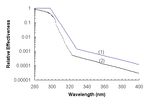
Figure 2 – (1) Human erythema action spectrum (CIE, 1987) and (2) actinic action spectrum (IRPA, 1989).
No action spectrum for melanoma in humans is available, however, Setlow et al. (1993) have measured the action spectrum for melanoma induction in Xiphophorus fish. As shown in Figure 3, this action spectrum has a relative effectiveness up to 800 times higher than that for erythema in the UVA waveband. Additionally, Figure 3 shows the action spectra for photoconjunctivitis (CIE, 1986a) and photokeratitis of the human eye (CIE, 1986b).
Several action spectra for various biological effects in plants are presented in Figure 3. The greatest influence of damaging biological effects is for wavelengths at about or below 300 nm, which is the waveband, where ozone depletion will have the greatest effect on UV irradiance. Caldwell (1971) provided a generalised plant damage action spectrum by incorporating the data from eight published spectra for UVC and UVB. This spectrum was formulated by Green et al. (1974) as:
Sl = 2.618[1.0-(l /313.3)2]e- (l -300)/31.08 for l < 313.3 nm (2.2)
with the action spectrum equal to zero for wavelengths greater than 313.3 nm. This action spectrum has been and is currently widely employed in research reported on the effects of UV on plants. A variety of photobiological responses in cucumbers has been measured to produce the median action spectrum plotted in Figure 3 (Coohill, 1989).
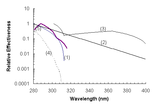
Figure 3 - Action spectra for (1) generalised plant damage (Caldwell, 1971) and (2) plant damage (Coohill, 1989) and the action spectra for (3) fish melanoma (Setlow, 1993) and (4) photoconjunctivitis (CIE, 1986a) and (5) photokeratitis (CIE, 1986b) of the human eye.
3. Biologically Effective Irradiance
3.1 Introduction
The degree of absorption of radiation by important biological macromolecules is wavelength dependent and as a result, the interfering effect of UV radiation is wavelength specific. Consequently, it is necessary to weight the incident spectral irradiance with a function called the action spectrum, Sl , which expresses the relative effectiveness of UV radiation at various wavelengths for a particular biological process (Caldwell et al., 1986). For a selected biological process, the biologically effective UV irradiance (Eeff ) is written as:
![]() (3.1)
(3.1)
where El is the spectral irradiance in W m-2 nm-1 and D l is the wavelength step in nm for the measurements of the spectral irradiance. The summation interval is the wavelength range from 200 nm to 400 nm. If the waveband consideration includes the vacuum ultraviolet region (< 200 nm) then the lower limit can be extended. The biologically effective UV irradiance has units of irradiance, namely, W m-2.
3.2 Comparison
Action spectra are necessary in assessing the biological effects of changes in the spectral irradiance due to changes in ozone concentrations. Additionally, they are necessary in relating the biologically effective dose from solar UV and spectrally different artificial sources (Caldwell et al., 1995). The three action spectra (Figure 4), namely, for photoconjunctivitis of the human eye, human erythema and plant damage have been selected as examples. The action spectrum for photoconjunctivitis has a negligible response in the UVA waveband. The response of the erythemal action spectrum in the UVA waveband is at least 3 decades lower than that in the UVB waveband. On the other hand, the response of the plant damage in the UVA waveband is significant as compared with that in the UVB waveband. The spectral biologically effective irradiances for a solar spectrum in autumn have been calculated for each of the three action spectra and are plotted on the second axis in Figure 4. The biologically effective irradiances calculated for these action spectra are provided in Table 1. The percentage contributions from the UVA and UVB wavebands are dependent on the action spectrum. For photoconjunctivitis of the human eye, there is no contribution from the UVA waveband, whereas, for plant damage, there is a 79% contribution for this solar spectrum. This is due to the relatively high effectiveness of this action spectrum in the UVA and the higher solar spectral irradiances in the UVA compared to the UVB. The percentage contribution of the UVA waveband for human erythema is 23% for this solar spectrum. This still forms an important contribution to the total human erythemal exposure.
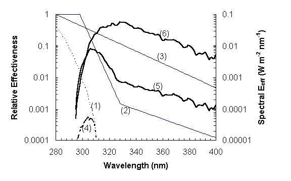
Figure 4 – Comparison of action spectra with the action spectrum for (1) photoconjunctivitis of the human eye, (2) human erythema and (3) plant damage and the resulting spectral biologically effective spectral irradiances for (4) photoconjunctivitis, (5) human erythema and (6) plant damage.
Table 1 - The biologically effective irradiances from a solar exposure to autumn sun and the percentage contributions from the UVA and UVB wavebands.
| Action Spectrum | Eeff | Percentage in the | Percentage in the |
| (W m-2) | UVB waveband | UVA waveband | |
| Photoconjunctivitis | 0.00052 | 100 | 0 |
| Human erythema | 0.15 | 77 | 23 |
| Plant damage | 2.36 | 21 | 79 |
4. Spectroradiometry
4.1 Introduction
Spectral irradiance measurements are more reliable than measurements with broadband instruments, as spectroradiometers can be more accurately calibrated to a standard lamp having its calibration traceable to the UV standard at the National Measurements Laboratory (Bernhard et al., 1997). One of the advantages of spectral irradiance measurements compared to broadband data is that they can be accurately weighted with the biological action spectrum for any biological process. Additionally, spectroradiometers are normally employed for the calibration of broadband instruments and dosimeters.
Accurate measurements of the UV spectral irradiance require a spectroradiometer with the minimum equipment of entrance optics with a diffuser or integrating sphere, monochromator, detector, amplifier and a control and acquisition unit (Seckmeyer et al., 1994). The system must have (Wong et al., 1995):
High wavelength resolution of usually 1nm;
Sensitivity of about 0.1 m W cm-2 at 300 nm;
High stray light rejection in the visible waveband;
Stable detector.
For solar measurements, additional requirements are necessary, namely:
Good cosine response;
Fast automatic scans:
Temperature stability and/or temperature compensation.
4.2 Spectroradiometer Intercomparisons
Spectroradiometers to measure UV spectral irradiance are complex and sensitive instruments and careful methodologies must be followed to maintain the reliability of the data (McKenzie et al., 1993). The instruments require wavelength calibration and calibration for absolute irradiance for each measurement session. Wavelength calibration is obtained by comparison with known spectral lines with further wavelength checking against the Fraunhofer absorption lines of the solar spectrum. Absolute intercomparisons of spectroradiometers have been performed both in the laboratory (Webb et al., 1994) and in the field (McKenzie et al., 1993, Seckmeyer et al., 1994a, 1995a, 1995b, Slaper et al., 1995, Gardiner et al., 1993) to assure the absolute and relative comparison of results. These intercomparisons found some instruments to intercompare within tolerances of ± 2%, others within ± 10%, with the differences for some instruments larger than this, particularly at wavelengths shorter than 300 nm. Additionally, the irradiance standards adopted must be traceable to the same standard (McKenzie et al., 1993). This process of intercomparison is lengthy and time consuming: however, other researchers have found that it is a necessary undertaking prior to comparisons of solar UV spectra measured at different sites with different instruments (Seckmeyer et al., 1995b). In order to eliminate some of the errors between spectroradiometers, researchers have developed methods for correcting for cosine errors (Seckmeyer and Bernhard, 1993, Feister et al., 1997) along with new entrance optics for spectroradiometers (Bernhard and Seckmeyer, 1997).
4.3 Spectral UV Measurements
Simultaneous spectroradiometer measurements at two different altitudes were undertaken in a Northern Hemisphere Alpine region where an increase of 24% at 300 nm between two sites separated vertically by 1000 m was measured (Blumthaler et al., 1994). The increase was wavelength dependent with an increase of 9% at 370 nm (Blumthaler et al., 1997). The UV climatology in the Southern Hemisphere is different from that in the Northern Hemisphere, with a series of spectroradiometer measurements finding the biologically effective UV to be approximately 40% higher in the mid southern latitudes compared to the corresponding northern latitudes (Seckmeyer et al., 1995b).
A series of spectral solar UV measurements were performed in tropical Australia with the average erythemal exposures exceeding those measured in Germany by between 55 and 70% (Bernhard et al., 1997). These differences are attributed to the smaller solar zenith angles and lower amounts of ozone in the atmospheric column in tropical Australia. Another set of spectroradiometer measurements at tropical latitudes is at the Mauna Loa Observatory where very high erythemal irradiances of 51 m W cm-2 have been recorded (Bodhaine et al., 1996, 1997). Additionally, spectroradiometer UV irradiances have been recorded at sub tropical latitudes (Wong et al., 1995) and at higher latitudes (McKenzie et al., 1992, Bais et al., 1993, Bittar and Mckenzie, 1990, Diaz et al., 1996, Webb, 1992, Seckmeyer et al., 1994b, Roy et al., 1997, Bojkov et al., 1995).
Total atmospheric ozone has been investigated with spectroradiometers with Roy et al., (1990) employing a spectroradiometer to measure anti-correlations between total atmospheric ozone and UVB irradiances. A method to derive total ozone from spectral measurements has been described by Stamnes et al., (1991). Spectral UV measurements have been employed to determine total atmospheric ozone with results within 1.1% and 0.4% of a Dobson and Brewer spectrophotometer respectively (Huber et al., 1995). McKenzie et al., (1991) established that variations in solar zenith angle and clouds have a higher relative importance on the erythemal irradiances than variations in ozone.
5. Broadband Measurements
5.1 Broadband Radiometers
Broadband radiometers measure the total irradiance over a selected waveband. The biologically effective irradiance can be determined if suitable filters and sensors are selected to provide a spectral response similar to the action spectrum in consideration.
The requirement permits a satisfactory calibration of the meter for applications in the field where the variation of the source spectrum is expected.
Perhaps the most popular instrument is the Robertson-Berger meter for the measurement of the erythemally effective irradiance. The device is based on (Berger, 1976) the use of a filter and a sensor (magnesium tungstate MgWO4) for the measurement of erythema UV. It is manufactured by the Solar Light Co. The instrument comes in different formats including one version with an electronic circuit for integrated exposures. It is reasonably accurate for measurements of artificial light sources provided the meter is calibrated against the source spectrum prior to the measurement. The sensitivity of the RB meter is about 0.56 m W cm-2 and polysulphone can record an exposure of about 2 mJ cm-2. For solar measurements, the temperature of the sensor must be maintained at a constant level. The model (501) known as the UV-Biometer (Wengraitis et al., 1998) is weatherproofed and temperature stabilised. It can be used for long-term field measurements of solar UV. Other manufacturers such as the International Light and the Biospherical Instrument also provide various models of UV meters. An intrinsic problem with the broadband UV meters is the difference between the spectral response of the instrument and the action spectrum in consideration. Furthermore, unlike spectroradiometers, broadband meters are usually fitted with a quartz diffuser instead of an integrating sphere. The cumulative error of the spectral response and the cosine response may be up to 11% over one month (Wengraitis et al., 1998) for measurements of solar radiation.
5.2 Passive Dosimeters
Chemical materials such as some plastics change when sunlight is applied to the materials. By measuring the degree of the change, information about the solar exposure can be deduced. A material suitable to be an ultraviolet dosimeter for outdoor measurements must have the following properties:
A spectral response similar to the action spectrum of the biological process in consideration;
Good cosine response;
Minimal temperature effects;
Rugged and portable.
Several detectors (Davis et al., 1976, Diffey, 1989, Wong et al., 1989) have been developed for different purposes. One of them – polysulphone, has been used extensively for measurements of human exposure to skin and it has been calibrated for the erythemally effective exposure (for example, Airey et al., 1997, Kimlin et al., 1998).
Biological materials can be used for assessing UV exposure. Since the material is biological specimens, its spectral response follows that of other biological bodies. An example is the utilisation of spore films for the measurement (Horneck et al., 1993). Studies (Quintern et al., 1997) show that the uncertainties of this type of detectors are comparable to that of chemical detectors. The dosimetric service is provided by the BioSense under the trade name VioSpor.
6. Spectrum Evaluator
Calibrated spectroradiometers may be utilised to measure the spectral irradiances, however the equipment is expensive and impractical for all situations. For example, they may be impractical for the measurement of biologically effective exposures at multiple sites simultaneously over humans and plants. They are impractical, if not impossible for the assessment of erythemal UV exposure to specific human body sites caused by solar radiation filtered by glass or plastic in field situations when the solar UV irradiances may be low (about 0.01 m W cm-2), for example, in cars and greenhouses. A new and novel detector (Parisi et al., 1997), called a spectrum evaluator (SE) was developed for estimating the solar UV spectrum in these situations when a spectroradiometer may be impractical.
The device with an overall size of 3 cm x 3 cm is based on four different dosimeter materials, namely, polysulphone, nalidixic acid, 8-methoxypsorolen and phenothiazine (Parisi and Wong, 1996a). Each of the materials is mounted in the holder with four holes of 6 mm diameter with a different material in each of the holes. This places the dosimetric materials in close proximity to one another and ensures that the possible errors due to spatial variation of the spectrum are eliminated and this miniaturisation also allows the spectrum evaluator to be applied on objects with complicated topography such as humans and on plant canopies or even within a plant canopy. The method requires a number of suitable materials, each with a different spectral response with their combined responses covering the entire UV waveband. These materials in thin film form change their optical absorbance after exposure to ultraviolet radiation. Each type of film is responsive to different UV wavebands and as a result the spectral irradiance falling on the device can be numerically calculated from the change of absorbance induced by the ultraviolet radiation (Parisi et al., 1997).
The method has been tested for the evaluation of both solar UV spectra and lamp spectra. The largest difference between the irradiances calculated with the evaluated and measured spectra is not more than 20% (Parisi et al., 1997). The method does not detect any fine structure in the spectrum; the evaluated function is a smoothed version of the spectral irradiance. The biologically effective irradiance may also be calculated for a particular biological process using the appropriate action spectrum. For example, the biologically effective exposures to plants has been measured both in the greenhouse (Parisi et al., 1996, 1998a) and in the field (Parisi and Wong 1996b, Parisi et al., 1998b). The SE has been employed for measuring the erythemal exposure resulting from the UVA waveband of an artificial light source (Wong and Parisi, 1996) and to evaluate the erythemal exposure due to filtered solar UV. For filtered solar UV, it has been employed to measure the erythemal UV exposures in a small glass enclosure designed to simulate a larger scale structure (Parisi and Wong, 1997a) and the personal UV exposures due to filtered UV radiation in field situations, for example, in cars and in greenhouses (Parisi and Wong, 1997b, 1997c, 1998).
7. Practicality
7.1 Measurement of Spectral Irradiance
Commercially available spectroradiometers can only be used for measurements of ambient radiation. For exposure to micro-organisms, such as cell irradiation, this is a good method for assessing the UV effect. In order to estimate the exposure to a specific tissue of a large biological body such as the skin of the human body, a model calculation based on the ambient exposure is required. A properly calibrated spectroradiometer can be used to measure the spectral irradiance of an artificial source with an uncertainty less than 5%. For solar measurements, because of the effect of the temperature change, it is not possible to claim an accuracy better than 10% unless the instrument is fully temperature stabilised, otherwise, the instrument has to be re-calibrated before each experimental session which lasts for a few hours. An example of a supplier for spectroradiometers is the Optronic Laboratories, Inc., 4470 35TH St., Orlando, Florida USA. The stray light level of this spectroradiometer is less than 10-8 at 285 nm. The wavelength accuracy is between ± 0.1 to ± 0.3 nm with a repeatability of ± 0.05 nm and a half bandwidth from 1 to 20 nm.
It is possible to estimate the spectral irradiance at a specific site using a spectrum evaluator. The result can be used for assessment of the biological effect of UV with an accuracy of about 20%. The method is particularly useful when one wants to assess the effect of various types of biological processes.
7.2 Broadband Measurements
Commercially available radiometers come in different forms. Portable meters are usually operated with batteries and can be employed for on site measurements. Some examples are tabulated in Table 2. These meters will have to be calibrated against a spectroradiometer at least twice a year. The accuracy of these instruments ranges from about 5% to 20% depending on the frequency of calibration.
Table 2 - Broadband radiometers
| Model | Waveband | Temperature Stabilisation | Address of Supplier |
| UV-Biometer 501 | UVB, Erythemal response |
± 0.01 oC | Solar Light Co., Inc. 721 Oak Lane, Philadelphia, PA. 19126. |
| RB 5D meter | UVB, Erythemal response |
Nil | Solar Light Co., Inc. 721 Oak Lane, Philadelphia, PA. 19126. |
| IL 1700, 1400 meters | UVB, UVA and UVR, Sensor probe comes with different responses |
Nil | International Light, 17 Graf Road, Newburyport, Mass. 01950-4092, USA) |
| Monitor Sensor | UVB, UVA and UVR | Nil | Monitor Sensors, 7-9 Industry Drive, Caboolture, Queensland, Australia |
Dosimeters with proper calibration can be used to estimate the exposure. They are available in a form of film badges. The accuracy is about 20% if the dosimeter is calibrated for each season. A summary of some popular dosimeters is provided below in Table 3.
Table 3 – UV dosimeters.
| Material | Wave band | Badge Size (cm2) | Response | Suppliers |
| Polysulphone | UVB | 3 x 3 | Erythemal | Joe Wong or Alfio Parisi, authors of this paper |
| CR-39 | UVB | 1 x 1 | Erythemal | Pershore Moulding Co., UK. |
| Viospor | UVA, UVB | Disc, 3.2 cm dia. 0.9 cm thick |
Erythemal | BioSense, Laboratory for Biological Sensory Systems, Quintern & Holtschmidt GbR, Mornesrstr. 4, D-533332, Bornheim – Germany |
| Spectrum evaluator | UVR | 3 x 3 | UV | Alfio Parisi, author of this paper |
8. Conclusion
Measurements of UVR require planning based on the type of experiments and the accuracy. To determine the ambient exposure or the exposure to micro-organism, the measurement is best performed with a calibrated spectroradiometer. If the measurement is to be made outdoors, the spectroradiometer should have a sufficient temperature control device. Suitable temperature correction must be made in order to obtain the required accuracy. Calibration should be made frequently (at least every six months) in order to minimise the error caused by the drifting of electronics. This method will provide an accuracy of about 10%.
To assess personal exposure to a biological body, a broadband radiometer or a passive dosimeter may be used. The device must be calibrated against the source spectrum with a calibrated spectroradiometer prior to the measurement. The sensitivity of the Robertson-Berger meter is about 0.56 m W cm-2 and polysulphone can record an exposure of about 2 mJ cm-2. It is expected the result would be no better than 20% accurate. To minimise the error caused by the difference between the action spectrum and the spectral response of the detector, a spectrum evaluator can be applied to estimate the source spectrum. The device can be used to measure exposures where the actinic and erythemal action spectra differ significantly. It can also be used to assess exposure due to low levels of UV (about 0.01 m W cm-2) caused by radiation filtered through glasses or plastic. The error introduced by the last method is about 20%.
REFERENCES
Airey, D.K., Wong, J.C.F., Fleming, R.A. & Meldrum, L.R. 1997, "An estimate of the total UV-B exposure for outdoor workers during a south-east Queensland summer," Health Phys. 72(4), 544-549.
Anders, A., Altheide, H., Knalmann, M. & Tronnier, H. 1995, "Action spectrum for erythema in humans investigated with dye lasers," Photochem. Photobiol. 61(2), 200-205.
Bais, A.F., Zerefos, C.S., Meleti, C., Ziomas, I.C. & Tourpali, K. 1993, "Spectral measurements of solar UVB radiation and its relations to total ozone, SO2 and clouds," J. Geophys. Res. 98(D3), 5199-5204.
Berger, D. 1976, "The sunburning ultraviolet meter: design and performance," Photochem. Photobiol. 24, 587-593.
Bernhard, G., Mayer, B. & Seckmeyer, G. 1997, "Measurements of spectral solar UV irradiance in tropical Australia," J. Geophys. Res. 102(D7), 8719-8730.
Bernhard, & Seckmeyer, G. 1997, "New entrance optics for solar spectral UV measurements," Photochem. Photobiol. 65(6), 923-930.
Bittar, A. & McKenzie, R.L. 1990, "Spectral ultraviolet intensity measurements at 45oS:1980 and 1988," J. Geophys. Res. 95(D5), 5597-5603.
Blumthaler, M., Webb, A.R., Seckmeyer, G., Bais, A.F., Huber, M. & Mayer, B. 1994, "Simultaneous spectroradiometry: A study of solar UV irradiance at two altitudes," Geophys. Res. Lett. 21(25), 2805-2808.
Blumthaler, M., Ambach, W. & Ellinger, R. 1997, "Increase in solar UV radiation with altitude," J. Photochem. Photobiol B: Biology, 39, 130-134.
Bodhaine, B.A., Mckenzie, R.L., Johnston, P.V., Hofmann, D,J., Dutton, E.G., Schnell, R.C., Barnes, J.E., Ryan, S.C. & Kotkamp. M. 1996, "New ultraviolet spectroradiometer measurements at Mauna Loa Observatory," Geophys. Res. Lett. 23(16), 2121-2124.
Bodhaine, B.A., Dutton, E.G., Hofmann, D,J., Mckenzie, R.L., & Johnston, P.V., 1997, "UV measurements at Mauna Loa: July 1995 to July 1996," J. Geophys. Res. 102(D15), 19265-19273.
Bojkov, R.D., Fioletov, V.E. & Diaz, S.B. 1995, "The relationship between solar UV irradiance and total ozone from observations over southern Argentina," Geophys. Res. Lett. 22(10), 1249-1252.
Caldwell, M.M. 1971, "Solar ultraviolet radiation and the growth and development of higher plants," in Photophysiology, ed. A.C. Giese, vol.6, pp.131-177, Academic Press, New York.
Caldwell, M.M., Gold, W.G., Harris, G. & Ashurst, C.W. 1983, "A modulated lamp system for solar UV-B (280-320nm) supplemental studies in the field," Photochem. Photobiol. 37(4), 479-485.
Caldwell, M.M., Camp, L.B., Warner, C.W. & Flint, S.D. 1986, "Action spectra and their key role in assessing biological consequences of solar UV-B radiation," in Stratospspric Ozone Reduction, Solar Ultraviolet Radiation and Plant Life, eds. R.C. Worrest & M.M. Caldwell, pp.87-111, Springer, Heidelberg.
Caldwell, M.M., Teramura, A.H., Tevini, M., Bornman, J.F., Bjorn, L.O. & Kulandaivelu, G. 1995, "Effects of increased solar ultraviolet radiation on terrestrial plants," Ambio, 24(3), 166-173.
CIE (International Commission on Illumination) Research Note 1986a, Photoconjunctivitis, CIE J. 5, 24-28.
CIE (International Commission on Illumination) Research Note 1986b, Photokeratitis, CIE J. 5, 19-23.
CIE (International Commission on Illumination) Research Note 1987, A reference action spectrum for ultraviolet induced erythema in human skin, CIE J. 6, 17-22.
Churchill, M.E., Peak, J.G. & Peak, M.J. 1991, "Correlation between cell survival and DNA single-strand break repair proficiency in the Chinese hamster ovary cell lines AA8 and EM9 irradiated with 365 nm ultraviolet A radiation," Photochem. Photobiol. 53, 229-236.
Coohill, T.P. 1989, "Ultraviolet action spectra (280 to 380 nm) and solar effectiveness spectra for higher plants," Photochem. Photobiol. 50(4), 451-457.
Coohill, T.P. 1991, "Action spectra again?" Photochem. Photobiol. 54(5), 859-870.
Davis, A., Deane, G.H.W. & Diffey, B.L. 1976, "Possible dosimeter for ultraviolet radiation," Nature, 261, 169-170.
de Gruijl, F.R., Sterenborg, H.J.C.M., Forbes, P.D., Davies, R.E., Cole, C., Kelfkens, G., van Weelden, H., Slaper, H. & van der Leun, J.C. 1993, "Wavelength dependence of skin cancer induction by ultraviolet irradiation of albino hairless mice," Cancer Res. 53, 53-60.
Diaz, S.B., Frederick, J.E., Lucas, T., Booth, C.R. & Smolskaia, I. 1996, "Solar ultraviolet irradiance at Tierra del Fuego: Comparison of measurements and calculations over a full annual cycle," Geophys. Res. Lett. 23(4), 355-358.
Diffey, B.L. 1989, "Ultraviolet radiation dosimetry with polysulphone film", in Radiation Measurement in Photobiology, ed. B.L. Diffey, pp.136-159, Academic Press, New York.
Feister, U., Grewe, R. & Gericke, K. 1997, "A method for correction of cosine errors in measurements of spectral UV irradiance," Solar Energy, 60(6), 313-332.
Freeman, S.E., Gange, R.W., Sutherland, J.C., Matzinger, E.A. & Sutherland, B.M. 1987, "Production of pyrimidine dimers in human skin exposed in situ to UVA radiation," J. Invest. Dermatol. 91, 349-352.
Gardiner, B.G., Webb, A.R., Bais, A.F., Blumthaler, M, Dirmhirn, I., Forster, P., Gillotay, D., Henriksen, K., Huber, M., Kirsch, P.J., Simin, P.C., Svenoe, T., Weihs, P. & Zerefos, C.S. 1993, "European intercomparison of ultraviolet spectroradiometers," Environ. Tech. 14, 25-43.
Green, A.E.S., Sawada, T. & Shettle, E.P. 1974, "The middle ultraviolet reaching the ground," Photochem. Photobiol. 19, 251-259.
Hacham, H., Freeman, S.E., Gange, R.W., Maytum, D.J., Sutherland, J.C. & Sutherland, B.M. 1990, "Does exposure of human skin in situ to 385 or 405 nm UV induce pyrimidine dimers in DNA?" Photochemm. Photobiol. 52, 893-896.
Holmberg, M., Almassy, Zs, Langerberg, M. & Niejahr, B. 1985, "The repair of strand breaks in human lymphocytes exposed to near UV-radiaition (UVA) and far UV-radiation (UVC)," Photochem. Photobiol. 41, 437-444.
Horneck, G., Quintern, L.E., Tettberg, P., Eschweiler, U. & Gorgen, E. 1993, "Biological dosimetry of solar uv radiation," in Biologic Effects of Light 1993, ed. E. G. Jung and M. F. Holick, 501-506, Walter de Gruyter.
Huber, M., Blumthaler, M., Ambach, W. & Staehelin, J. 1995, "Total atmospheric ozone determined from spectral measurements of direct solar UV irradiance," Geophys. Res. Lett. 22(1), 53-56.
IRPA (International Radiation Protection Association) 1989, "Proposed change to the IRPA 1985 guidelines on limits of exposure to ultraviolet radiation," Health Phys. 56(6), 971-972.
Kimlin, M.G., Parisi, A.V. & Wong, J.C.F. 1998, "Quantification of the personal solar UV exposure of outdoor workers, indoor workers and adolescents at two locations in southeast Queensland," Photodermatol. Photoimmunol. Photomed. 14(1), 7-11.
Longstreth, J.D., de Gruijl, F.R., Kripke, M.L., Takizawa, Y. & van der Leun, J.C. 1995, "Effects of increased solar ultraviolet radiation on human health," Ambio, 24(3), 153-165.
McKenzie R.L., Matthews, W.A. & Johnston, P.V. 1991, "The relationship between erythemal UV and Ozone, derived from spectral irradiance measurements," Geophys. Res. Lett. 18(12), 2269-2272.
McKenzie R.L., Johnston, P.V., Kotkamp, M., Bittar, A. & Hamlin, J.D. 1992, "Solar ultraviolet spectroradiometry in New Zealand: instrumentation and sample results from 1990," Appl. Optics, 31, 6501-6509.
McKenzie R.L., Kotkamp, M., Seckmeyer, G., Erb, R., Roy, C.R., Gies, H.P. & Toomey, S.J. 1993, "First southern hemisphere intercomparison of measured solar UV spectra," Geophys. Res. Lett. 20(20), 2223-2226.
Parisi, A.V. & Wong, C.F. 1996a, "A new method for measurements of erythemal irradiance," Photodermatol. Photoimmunol. Photomed. 12(2), 171-179.
Parisi, A.V. & Wong, C.F. 1996b, "Plant canopy shape and the influences on UV exposures to the canopy," Photochem. Photobiol. 64(1), 143-148.
Parisi, A.V., Wong, C.F. & Galea, V. 1996, "A method for evaluation of UV and biologically effective exposures to plants," Photochem. Photobiol. 64(2), 326-333.
Parisi, A.V., Wong, C.F. & Moore, G.I. 1997, "Assessment of the exposure to biologically effective UV radiation using a dosimetric technique to evaluate the solar spectrum," Phys. Med. Biol. 42, 77-88.
Parisi, A.V. & Wong, C.F. 1997a, "Erythemal irradiances of filtered ultraviolet radiation," Phys. Med. Biol. 42(7), 1263-1275.
Parisi, A.V. & Wong, C.F. 1997b, "The erythemal ultraviolet exposure for humans in greenhouses," Phys. Med. Biol. 42(12), 2331-2339.
Parisi, A.V. & Wong, C.F. 1997c, "Human exposure to filtered solar ultraviolet radiation," Proceedings of the First Internet Conference on Photochemistry and Photobiology, Nov 17 -Dec 19 1997, Internet Photochemistry and Photobiology.
Parisi, A.V. & Wong, C.F. 1998, "Quantitative evaluation of the personal erythemal ultraviolet exposure in a car," Photodermatol. Photoimmunol. Photomed. 14(1), 12-16.
Parisi, A.V., Wong, C.F. & Galea, V. 1998a, "A study of the total ultraviolet exposure to all the leaves for small plant growth," J. Photochem. Photobiol. B: Biology, 45(1), 36-42.
Parisi, A.V., Wong, C.F. & Randall, C. 1998b, "Simultaneous assessment of photosynthetically active and ultraviolet solar radiation," Agricult. For. Meteorol. 92(2), pp.97-103.
Peak, J.G. & Peak, M.J. 1991, "Comparison of initial yields of DNA to protein crosslinks induced in human cells in culture by x-ray, gamma rays, JANUS 0.85 MeV neutrons, light photons in the UVC, UVB, UVA regions and blue light.," Mutat. Res. 246, 187-191.
Quaite, F.E., Sutherland, B.M. & Sutherland, J.C. 1992, "Action spectrum for DNA damage in alfalfa lowers predicted impact of ozone depletion," Nature, 358 (6387), 576-578.
Quintern, L.E., Furusawa, Y., Fukutsu, K. & Holtschmidt, H. 1997, "Characterization and application of uv detector spore films: the sensitivity curve of a new detector system provides good similarity to the action spectrum for uv-induced erythema in human skin," J. Photochem. Photobiol. B: Biology. 37, 158-166.
Roy, C.R., Gies, H.P. & Elliot, G. 1990, "Ozone Depletion," Nature, 347, 235-236.
Roy, C.R., Gies, H.P. & Toomey, S. 1997, "Monitoring UV-B at the Earth’s surface," Aust Met Mag. 46, 203-210.
Roza, L., van der Schans, G.P., & Lohman, P.H.M. 1985, "The induction and repair of DNA damage and its influence on cell death in primary human fibroblasts exposed to UV-A or UV-C radiation," Mutat. Res. 217, 219-226.
Seckmeyer, G. & Bernhard, G. 1993, "Cosine error correction of spectral UV-irradiances," SPIE Proc. Bellingham, Washington, 2049, 140-151.
Seckmeyer, G., Thiel, S., Blumthaler, M., Fabian, P., Gerber, S., Gugg-Helminger, A., Hader, D.P., Huber, M., Kettner, C., Kohler, U., Kopke, P., Maier, H., Schafer, J., Suppan, P., Tamm, E. & Thomalla, E. 1994a, "Intercomparison of spectral UV radiation measurement systems," Appl. Optics, 33(33), 7805-7811.
Seckmeyer, G., Mayer, B., Erb, R. & Bernhard, G. 1994b, "UV-B in Germany higher in 1993 than in 1992," Geophys. Res. Lett. 21(7), 577-580.
Seckmeyer, G., Bernhard, G., Mayer, B. & Erb, R. 1995a, "High-accuracy spectroradiometry of solar ultraviolet radiation," Metrologia, 32, 697-700.
Seckmeyer, G., Mayer, B., Bernhard, G., McKenzie, R.L., Johnston, P.V., Kotkamp, M., Booth, C.R., Lucas, T., Mestechkina, T., Roy, C.R., Gies, H.P. & Tomlinson, D. 1995b, "Geographical differences in the UV measured by intercompared spectroradiometers," Geophys. Res. Lett. 22(14), 1889-1892.
Setlow, R.B. 1974, "The wavelengths of sunlight effective in producing skin cancer: a theoretical analysis," Proc Natl. Acad. Sci. USA, 71, 3363-3366.
Setlow, R.B., Grist, E., Thompson, K. & Woodhead, A.P. 1993, "Wavelengths effective in induction of malignant melanoma," Proc. Natl. Acad. Sci. 90, 6666-6670.
Sharma, R.C. & Smith, K.C. 1985, "A mechanism for rich-medium inhibition of the repair of daughter-strand gaps in the deoxyribonucleic acid of UV-irradiated Escherichia coli K12 uvrA," Mutat. Res. 146, 177-183.
Slaper, H., Reinen, H.A.J.M., Blumthaler, M., Huber, M. & Kuik, F. 1995, "Comparing ground-level spectrally resolved solar UV measurements using various instruments: A technique resolving effects of wavelength shift and slit width," Geophys. Res. Lett. 22(20), 2721-2724.
Stamnes, K., Slusser, J. & Bowen, M. 1991, "Derivation of total ozone abundance and cloud effects from spectral irradiance measurements," Appl. Optics, 30(30), 4418-4426.
Tyrrell, R.M. & Pidoux, M. 1989, "Singlet oxygen involvement in the inactivation of cultured human fibroblasts by UVA (334 nm, 365 nm) and near-visible (405 nm) radiations," Photochem. Photobiol. 49, 407-412.
Webb, A.R. 1992, "Spectral measurements of solar ultraviolet-B radiation in Southeast England," J. App. Met. 31, 212-216.
Webb, A.R., Gardiner, B.G., Blumthaler, M., Forster, P., Huber, M. & Kirsch, P.J. 1994, "A laboratory investigation of two ultraviolet spectroradiometers," Photochem. Photobiol. 60(1), 84-90.
Wengraitis, S., Benedetta, D. & Sliney, D.H. 1998, "Intercomparison of effective erythemal irradiance measurements from two types of broad-band instruments during june 1995," Photochem. Photobiol. 68(2), 179-182.
Wong, C.F., Fleming, R. & Carter, S.J. 1989, "A new dosimeter for ultraviolet-B radiation," Photochem. Photobiol. 50(5), 611-615.
Wong, C.F. & Parisi, A.V. 1996, "Measurement of UVA exposure to solar radiation," Photochem. Photobiol. 63(6), 807-810.
Wong, C.F., Toomey, S., Fleming, R.A. & Thomas, B.W. 1995, "UV-B radiometry and dosimetry for solar measurements," Health Phys. 68(2), 175-184.