| Papers and Posters | Site Home Page |
8-METHOXYPSORALEN PLUS UVA INHIBITS TH1 CELL DERIVED INTERFERON-GAMMA AND INTERLEUKIN-2 BUT INDUCES TH2 CELL DERIVED INTERLEUKIN-4 EXPRESSION
Gabriele Klosner, Robert Knobler, Franz Trautinger and Peter Neuner.
Department of Dermatology, Division of Special and Environmental Dermatology,
University of Vienna, Währinger Gürtel 18-20, A-1090 Vienna, Austria. e-mail:
gabriele.klosner@akh-wien.ac.at
ABSTRACT
Treatment with 8-methoxypsoralen (8M0P) plus UVA (PUVA) has been shown to be beneficial in
inflammatory as well as in malignant skin disorders such as psoriasis, graft versus host
disease or cutaneous T-cell lymphomas. Since the ratio between interferon-gamma (IFNg)
producing TH1 and interleukin-4 (IL-4) producing TH2 cells appears to be critical for the
progression of skin disorders, the effect of PUVA on TH1- and TH2-cytokine production was
investigated.
Peripheral blood lymphocytes (PBL) derived from 6 healthy volunteers were stimulated
and treated as described in material and methods. FACS
analysis revealed undetectable amounts of INFg and IL-2 in unstimulated CD4+ cells.
However, upon stimulation with PMA and iononmycin, CD4+ cells showed a distinct production
of IFNg and IL-2 that was significantly downregulated by PUVA treatment. IL-4 production
was detected in unstimulated as well as in stimulated cells. In contrast to IFNg and IL-2,
PUVA treatment significantly upregulated IL-4 production.
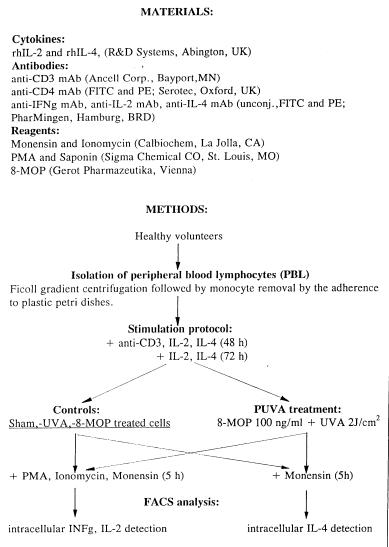
FIGURE 1, 2 AND 3:
Intracellular INFg, IL-2 and IL-4 production by PBL. PBL were stimulated with anti-CD3 mAb,
IL-2 and IL-4 for 48 hours, washed and subsequently stimulated with IL-2 and IL-4 for
further 72 hours. After washing, cells were incubated in the presence of 8-MOP (100 ng/ml)
for 1 hour and subsequently irradiated with UVA (2J/cm˛). They were stimulated with PMA,
ionomycin and monensin for further 5 hours.
Control cells were treated with 8-MOP, UVA alone, or were left untreated (control).
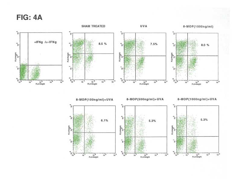
A: FACS analysis represents simultanous staining of PBL with anti-CD4 mAb (APC or PerCP, abscissa) and anti IFNg, anti IL-2 and anti IL-4 mAb (FITC or PE; ordinate), respectively.
B: Results are given as % of cells positive for the respektive cytokine (as analyzed by flow cytometry), compared to the number of cytokine producing cells in the control determined as 100%.
Paired t-test: (control vs PUVA, n=3) INFg: p=0.06, IL-2: p=0.06, IL-4: p=0.009
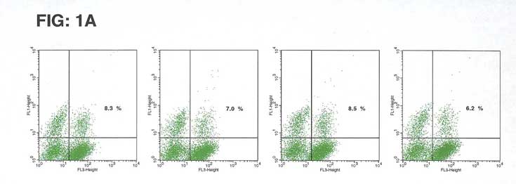
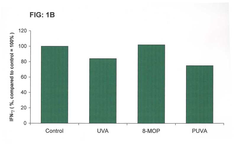
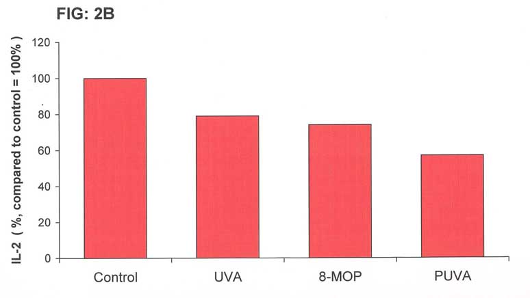
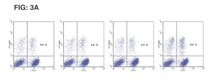
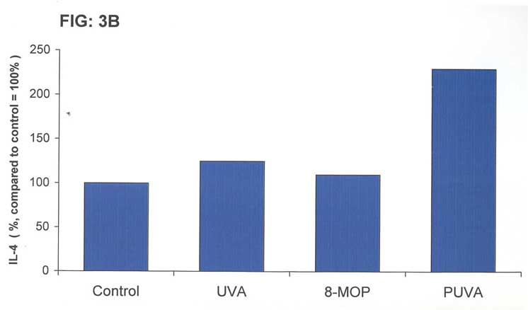
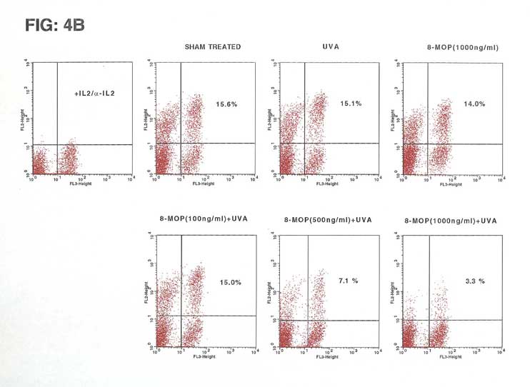
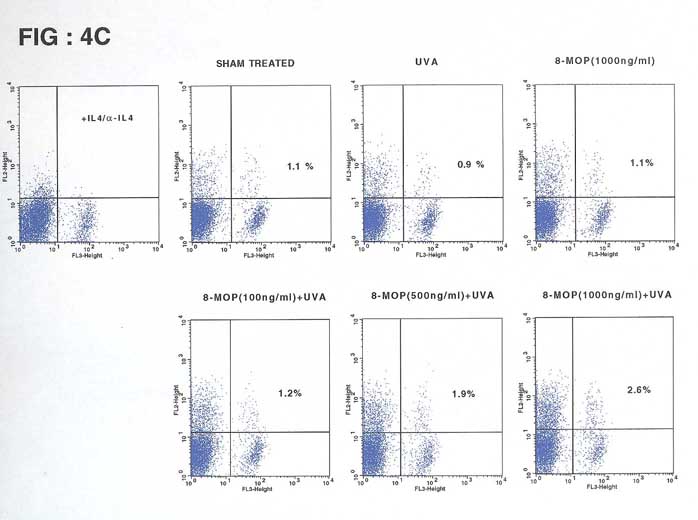
FIGURE 4 A, B and C:
Cells were cultured as described above, exposed to increasing doses of 8-MOP (100, 500 and 1000 ng/ml) plus UVA (2 J/cm˛) and tested for IFNg, IL-2 and IL-4
RESULTS AND CONCLUSION
These data show that PUVA treatment in vitro downregulates the expression of THl
derived cytokines such as IFNg and IL-2 and upregulates TH2 derived IL-4 in CD4+cells. lt
has to be determined whether the induction of a shift from TH1 to TH2 cytokine profile
plays a role in the response to photochemotherapy in inflammatory and malignant skin
diseases such as psoriasis, atopic dermatits, and cutaneous T-cell lymphoma.